Mid-Atlantic Division
Previously Offered (depleted) Tissue Microarrays
- CHTN2002N1 - Normal Human Tissues
- CHTN2002T1 - "Test" TMA
- CHTN2003CRCprog - Colorectal Carcinoma Progression
- CHTN BrCaProg1 - Breast Cancer Progression
- CHTN_BrCaProg2 - Breast Cancer Progression (Histologic Progression)
- CHTN OvCa1 - Ovarian Carcinoma Survey
- CHTN Test2 - Test Tissue Microarray
- CDP Breast Cancer Progression Tissue Microarray (Clinical Progression)
- CHTN_Norm2 - Normal Tissue Survey
- CHTN CNS1 - Human Nervous System
-
CHTN2002N1 - Normal Human Tissues
TMA series CHTN2002N1: Purpose: To provide researchers with a tissue microarray of formalin fixed paraffin embedded samples that includes most of the cell types present in the human body. Most samples are of non-neoplastic adult tissue obtained from surgical resection specimens, fixed within one hour of removal from the donors. The fixative used is buffered zinc formaldehyde (3.7% formaldehyde) (Z-fix, Anatech, LTD., Battle Creek, MI). Exceptions: Parathyoid - tissue is from a benign parathyroid adenoma due to the limiting size and limited availability of normal parathyroid tissue. Lymphatics-tissue is from a benign lymphangioma, due to the difficulty in sampling normal lymphatics Central nervous system tissue - all obtained from autopsy specimens within 36 hours of death. TMA design: Each tissue type was sampled multiple times with 0.6 mm needle cores in the original array design. The CHTN2002N1 TMA series was constructed as 4 replicate TMA blocks, designated CHTN2002N1A, CHTN2002N1B, CHTN2002N1C, CHTN2002N1D
Unstained histologic sections are 4 microns thick, on charged glass slides.
- Guide Sheets
- Download Guide Sheets (MS Excel)
- Download information sheet (pdf)
-
CHTN2002T1 - "Test" TMA
TMA series CHTN2002T1 contains 5 human tissue types (colonic mucosa, endometrium, spleen, liver and uterine smooth muscle) in 0.6 mm and 2 mm spot sizes. These are useful for determining assay conditions and procedures before using more expensive comprehensiveTMA slides.
Unstained histologic sections are 4 microns thick, on charged glass slides.
- Guide Sheets
- Download Guide Sheets (MS Excel)
- Download information sheet (pdf)
-
CHTN2003CRCprog - Colorectal Carcinoma Progression
The CHTN2003CRCprog TMA contains up to 20 cases of non-neoplastic colonic mucosa, 14 cases of adenomatous polyps, 14 cases of primary colorectal adenocarcinomas, 7 cases of adenocarcinoma metastatic to regional lymph nodes and 7 cases of adenocarcinoma metastatic to distant sites. Each case is sampled three times with 0.6 mm cores.
This TMA represents a limited number of cases that may detect strong trends in differential gene expression, and is intended for pilot surveys and generation of hypotheses. This TMA does not contain case numbers of sufficient quantity to prove the clinical utility of a marker.
Unstained histologic sections are 4 microns thick, on charged glass slides.
- Guide Sheets
- Download Guide Sheets (MS Excel)
- Download information sheet (pdf)
-
CHTN BrCaProg1 - Breast Cancer Progression
The CHTNBrCaProg1 TMA contains non-neoplastic breast tissue, ductal carcinoma in situ (DCIS), infiltrating ductal carcinoma of the breast, infiltrating lobular carcinoma of the breast, and cases of adenocarcinoma metastatic to regional lymph nodes. Each case is sampled one time with a 2 mm core. The TMA design includes 14 cases of non-neoplastic breast, 14 cases of DCIS, 21 cases of primary carcinoma and 7 cases of metastatic carcinoma. Histologic quality assurance limits ensure a minimum of 7 cases of non-neoplastic breast, 10 cases of infiltrating carcinoma primary to the breast and 4 cases of metatstatic carcinoma. Due to the difficulty in capturing pre-invasive neoplasia, a minimum number of DCIS cases is not being guaranteed.
This TMA represents a limited number of cases that may detect strong trends in differential gene expression, and is intended for pilot surveys and generation of hypotheses. This TMA does not contain case numbers of sufficient quantity to prove the clinical utility of a marker.
Unstained histologic sections are 4 microns thick, on charged glass slides.
- Guide Sheets
- Download Guide Sheets (MS Excel)
- Download information sheet (pdf)
-
CHTN_BrCaProg2 - Breast Cancer Progression (Histologic Progression)
The CHTN_BrCaProg2 TMA contains non-neoplastic breast tissue, ductal carcinoma in situ (DCIS), infiltrating ductal carcinoma of the breast, infiltrating lobular carcinoma of the breast, and cases of adenocarcinoma metastatic to regional lymph nodes. Each case is sampled one time with a 2 mm core. The TMA design includes 14 cases of non-neoplastic breast, 14 cases of DCIS, 21 cases of primary carcinoma and 7 cases of metastatic carcinoma. Histologic quality assurance limits ensure a minimum of 7 cases of non-neoplastic breast, 15 cases of infiltrating carcinoma primary to the breast and 4 cases of metatstatic carcinoma. Due to the difficulty in capturing pre-invasive neoplasia, a minimum number of DCIS cases is not being guaranteed.
This TMA represents a limited number of cases that may detect strong trends in differential gene expression, and is intended for pilot surveys and generation of hypotheses. This TMA does not contain case numbers of sufficient quantity to prove the clinical utility of a marker.
Unstained histologic sections are 4 microns thick on charged glass slides.
- View TMA Details (Maps & Tissue Info)
- Download information sheet (pdf)
- Download Guide Sheets (MS Excel)
-
CHTN OvCa1 - Ovarian Carcinoma Survey
The CHTN OvCa1 TMA contains examples of the major histologic types of epithelial ovarian cancer . Each case is sampled four times with 0.6 mm cores. The TMA design includes 20 cases each of serous papillary carcinoma (well to moderately differentiated), clear cell carcinoma, endometrioid adenocarcinoma and mucinous adenocarcinoma). Another 20 cases of poorly and undifferentiated carcinoma are also provided.
This TMA may detect strong trends in differential gene expression among the different histologic types of ovarian carcinoma, and is intended for pilot surveys and generation of hypotheses. This TMA is not designed to prove the clinical utility of prognostic biomarkers.
Unstained histologic sections are 4 microns thick on charged glass slides.
- View TMA Details (Maps & Tissue Info)
- Download information sheet (pdf)
- Download Guide Sheets (MS Excel)
-
CHTN Test2 - Test Tissue Microarray
CHTN Test Tissue Microarrays (versions 2,3,4) contain lymphoid, epithelial and stromal tissue (spleen, colonic mucosa, prostate, liver, breast cancer and uterine smooth muscle) in 0.6 mm spot sizes. These are useful for determining assay conditions and procedures before using more expensive comprehensiveTMA slides.
Unstained histologic sections are 4 microns thick on charged glass slides.
- View TMA Details (Maps & Tissue Info)
- Download information sheet (pdf)
- Download Guide Sheets (MS Excel)
-
CDP Breast Cancer Progression Tissue Microarray (Clinical Progression)
CDP Breast Cancer Progression Tissue Microarrays are designed to investigate differences in marker prevalence in three stages of invasive breast cancer: node-negative, node-positive and metastatic disease. This TMA set contains a total of 339 breast tissue cores, plus cell line and normal non-breast tissue controls. All of the invasive cases are primary breast cancers with a principal histology of ductal cancer. The CDP breast cancer progression TMA was designed by a National Cancer Institute biostatistician to ensure high statistical power for studies of stage specific markers of breast cancer.
Unstained histologic sections are 4 microns thick on charged glass slides. 1 set consists of 6 TMA slides.
- View TMA Details (Design & Tissue Info)
- View and Download TMA Annotations (Data Element Info, Complete Annotation Data, Maps)
- Download IHC & FISH staining info (pdf)
-
CHTN_Norm2 - Normal Human Tissues
The normal tissue TMA series contains 50 human tissue types in 1.0 mm spot sizes. Most samples are of normal, non-neoplastic adult tissue obtained from surgical resection specimens, excepting parathyroid from a hyperplastic parathyroid gland and central nervous system tissue (cerebral cortex and white matter) from autopsy.
This TMA is designed provide researchers with a screening tool containing most of the cell types present in the human body.
Unstained histologic sections are 4 microns thick on charged glass slides.
-
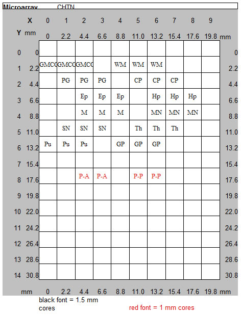
CHTN CNS1 - Human Nervous System
The CHTN CNS1 TMA samples 12 areas of the central nervous system and supporting structures, capturing most of the major cell types of the brain and spinal cord. Each target tissue type is sampled three times with 1.5 mm cores.
This TMA is meant to provide an initial survey tool for determining gene expression patterns in different cell types of the central nervous system.
Unstained histologic sections are 4 microns thick on charged glass slides.
- View TMA Details (Maps & Tissue Info)
- Download information sheet (pdf)
- Download Guide Sheets (MS Excel)