Mid-Atlantic Division
Currently Available Tissue Microarrays
The CHTN distributes TMAs of its own design and from allied organizations. The TMAs listed below with the “CDP” annotation were designed by the Cancer Diagnosis Program of the National Cancer Institute. The application process is slightly different for CDP TMAs than for CHTN TMAs, and the fees are more significant to reflect the larger number of cases, the greater degree of annotated data and the statistical design process associated with these resources. Please see instructions on how to request TMA slides.
Custom tissue microarray construction is not available as a CHTN service. However, the CHTN Mid-Atlantic Division is able to offer construction of TMAs utilizing specimen blocks provided by the Investigator on a fee-for-service basis in partnership with the UVA Biorepository & Tissue Research Facility. Please contact the CHTN Mid-Atlantic Division for further information.
Cooperative Human Tissue Network (CHTN) Cancer Tissue Microarrays
- CHTN_CRC2 - Colorectal Carcinoma Progression
- CHTN OvCa2 - Ovarian Carcinoma Survey
- CHTN_BrCaProg3 - Breast Cancer Progression (Histologic Progression)
- CHTN_BrCaStg1-Breast Cancer Progression (Clinical Progression)
- CHTN_PrCProg1 - Prostate Cancer Tumor Progression
- CHTN_PancProg1 - Pancreas Carcinoma Progression
- CHTN_PanCA1 - Pancreas Neoplasia (Survey)
- CHTN_Lung_AdenoCA1 - Lung Adenocarcinoma
- CHTN_Lung_SCC_Misc1 - Lung Squamous Cell and Misc Carcinoma
- CHTN_GBM1 - Glioblastoma Multiforme
Cooperative Human Tissue Network (CHTN) Normal and Test Tissue Microarrays
- CHTN_Norm3 - Normal Tissue Survey
- CHTNEndo1 - Normal Endometrial Cycle
- CHTN CNS2 - Human Nervous System
- CHTN Test3 - Test Tissue Microarray
Cancer Diagnosis Program (CDP) Tissue Microarrays
Special application process. More information here.
- CDP Breast Cancer Stage I Prognostic Tissue Microarray
- CDP Breast Cancer Stage II Prognostic Tissue Microarray
- CDP Breast Cancer Stage III Prognostic Tissue Microarray
- CDP Melanoma Progression Tissue Microarray
Cooperative Human Tissue Network (CHTN) Cancer Tissue Microarrays
-
CHTN_CRC2 - Colorectal Carcinoma Progression
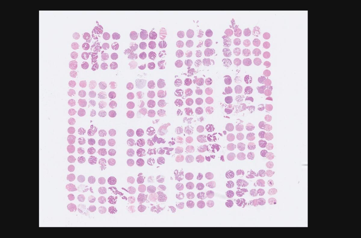
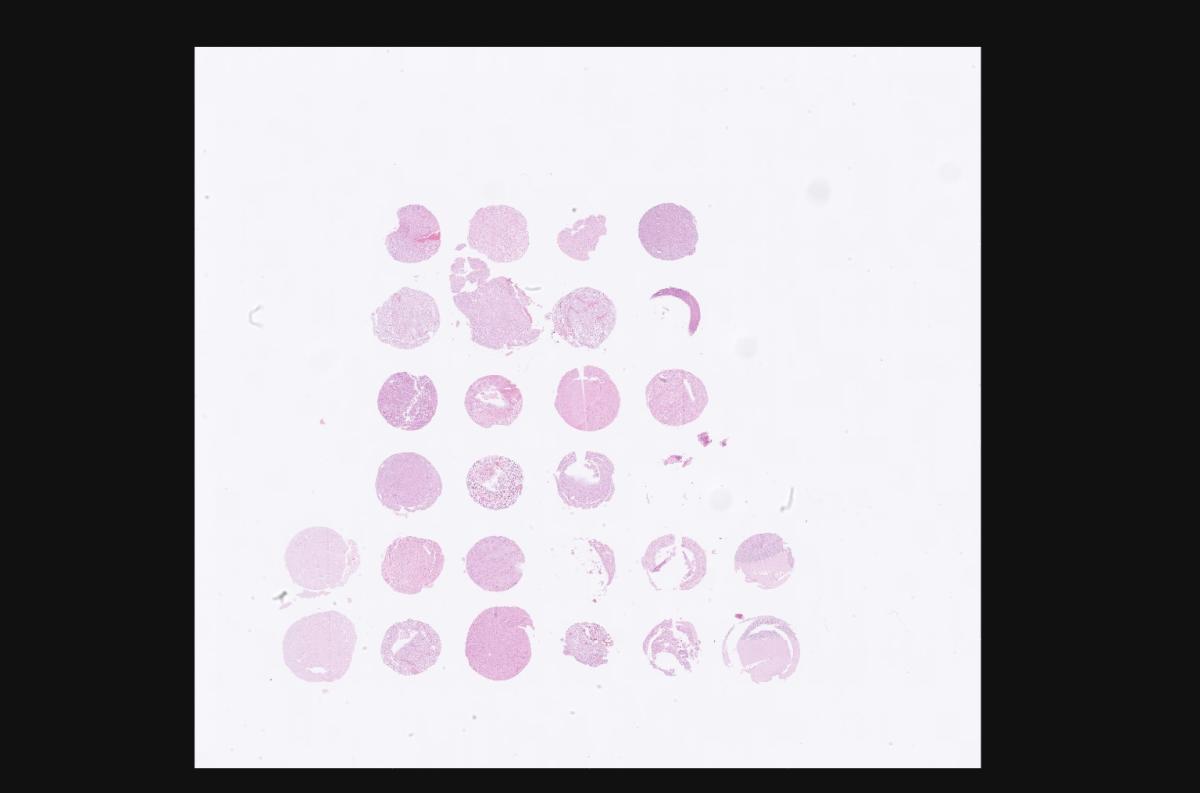
The CHTN_CRC2 TMA contains up to 20 cases of non-neoplastic colonic mucosa, 14 cases of adenomatous polyps, 14 cases of primary colorectal adenocarcinomas, 7 cases of adenocarcinoma metastatic to regional lymph nodes and 7 cases of adenocarcinoma metastatic to distant organs. Each case is sampled three times with 0.6 mm cores.
This TMA may detect strong trends in differential gene expression and is intended for pilot surveys and generation of hypotheses. This TMA is not designed to prove the clinical utility of prognostic biomarkers.
Unstained histologic sections are 4 microns thick on charged glass slides. A high resolution digital pathology H&E of the TMA is available upon request.
- Pricing:
Limit of 10 slides or slide sets per investigator
- Academic investigators: $75.00/slide plus shipping
- Commercial: $225.00/slide plus shipping
- International outside North America: $225.00/slide plus shipping
- View TMA Details (Maps & Tissue Info)
- Download information sheet (pdf)
- Download Guide Sheets (MS Excel)
- TMA Digital Image
- Pricing:
-
CHTN OvCa2 - Ovarian Carcinoma Survey
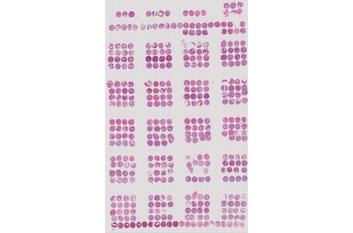
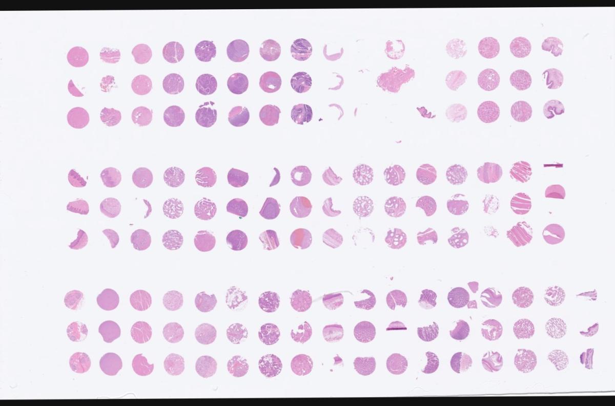
The CHTN OvCa2 TMA contains examples of human ovarian carcinoma and ovarian borderline tumors representing the major histologic types of epithelial ovarian tumors. Each case is sampled four times with 0.6 mm cores. The TMA design includes 12 cases each of serous papillary carcinoma, clear cell carcinoma, endometrioid adenocarcinoma and mucinous adenocarcinoma. Another 6 cases each of serous and mucinous borderline tumors are provided. In addition 24 cases of non-malignant tissues (proliferative endometrium, fallopian tube, serous and mucinous cystadenomas) are represented.
This TMA may detect strong trends in differential gene expression among the different histologic types of ovarian carcinoma, and is intended for pilot surveys and generation of hypotheses. This TMA is not designed to prove the clinical utility of prognostic biomarkers.
Unstained histologic sections are 4 microns thick on charged glass slides. A high resolution digital pathology H&E of the TMA is available upon request.
- Pricing:
Limit of 10 slides or slide sets per investigator
- Academic Investigators: $75.00/slide plus shipping
- Commercial: $225.00/slide plus shipping
- International outside North America: $225.00/slide plus shipping
- View TMA Details (Maps & Tissue Info)
- Download information sheet (pdf)
- Download Guide Sheets (MS Excel)
- TMA Digital Image
- Pricing:
-
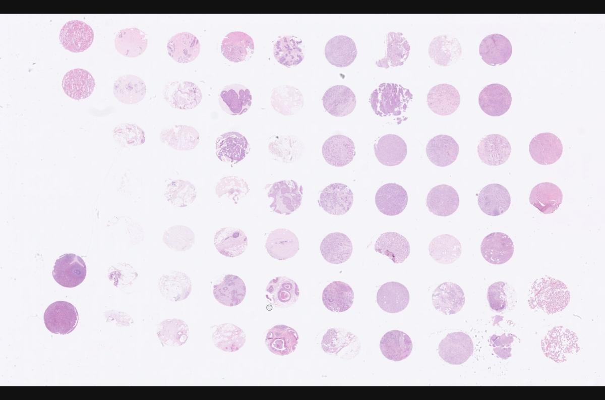
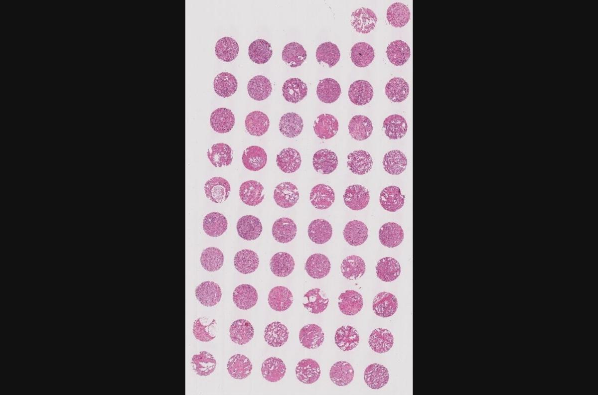
CHTN_BrCaProg3 - Breast Cancer Progression (Histologic Progression)
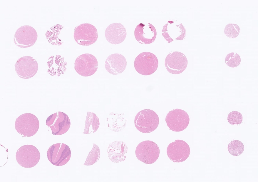
The CHTN_BrCaProg3 TMA contains non-neoplastic breast tissue, ductal carcinoma in situ (DCIS), ductal carcinoma of the breast, lobular carcinoma of the breast, and cases of breast carcinoma metastatic to lymph nodes. Each case is sampled one time with a 2 mm core. The TMA design includes 14 cases of non-neoplastic breast, 14 cases of DCIS, 21 cases of primary carcinoma and 7 cases of metastatic carcinoma. Histologic quality assurance limits ensure a minimum of 7 cases of non-neoplastic breast, 15 cases of infiltrating carcinoma primary to the breast and 4 cases of metastatic carcinoma. Due to the difficulty in capturing pre-invasive neoplasia, a minimum number of DCIS cases is not being guaranteed.
This TMA represents a limited number of cases that may detect strong trends in differential gene expression, and is intended for pilot surveys and generation of hypotheses. This TMA does not contain case numbers of sufficient quantity to prove the clinical utility of a marker.
Unstained histologic sections are 4 microns thick on charged glass slides. A high resolution digital pathology H&E of the TMA is available upon request.
- Pricing:
Limit of 10 slides or slide sets per investigator
- Academic investigators: $75.00/slide plus shipping
- Commercial: $225.00/slide plus shipping
- International outside North America: $225.00/slide plus shipping
- View TMA Details (Maps & Tissue Info)
- Download information sheet (pdf)
- Download Guide Sheets (MS Excel)
- TMA Digital Image
- Pricing:
-
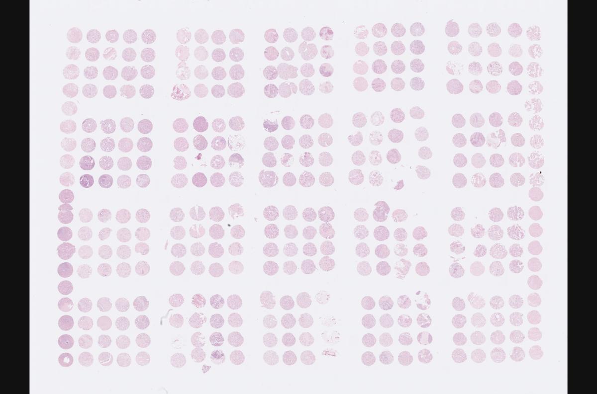
CHTN_BrCaStg1 - Breast Cancer Progression (Clinical Progression)
The CHTN BrCaStg1 TMA contains a variety of tumor stages (T1-T4) and provides clinical follow-up data. The TMA design includes 60 cases of primary node-negative invasive breast carcinoma, 60 cases of primary node-positive invasive breast carcinoma, 20 cases of breast carcinoma metastatic to local lymph nodes and 20 cases of breast carcinoma metastatic to distant sites.
This TMA represents a limited number of cases that may detect strong trends in differential gene expression, and is intended for pilot surveys and generation of hypotheses. This TMA does not contain case numbers of sufficient quantity to prove the clinical utility of a marker.
Unstained histologic sections are 4 microns thick on charged glass slides. A high resolution digital pathology H&E of the TMA is available upon request.
- Pricing:
Limit of 10 slides or slide sets per investigator
- Academic investigators: $250.00/slide plus shipping
- Commercial: $750.00/slide plus shipping
- International outside North America: $750.00/slide plus shipping
- View TMA Details (Maps & Tissue Info)
- Download information sheet (pdf)
- Download Guide Sheets (MS Excel)
- TMA Digital Image Block 1 | TMA Digital Image Block 2
- Pricing:
-
CHTN_PrC_Prog1 - Prostate Cancer Tumor Progression
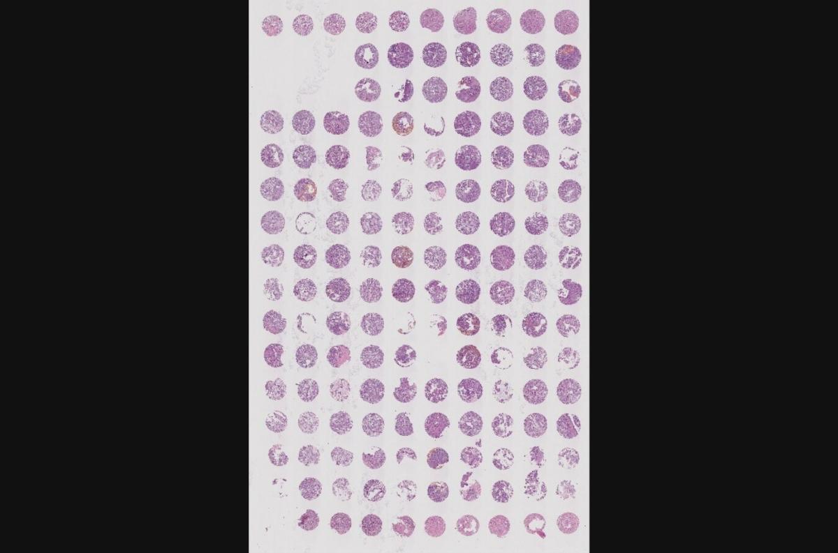
The CHTN_PrC_Prog1 TMA contains examples of prostate epithelium representing the stages of tumor progression in prostate adenocarcinoma. Each case is sampled one time with a 1.5 mm core. The TMA design includes 10 cases each of non-neoplastic prostate glands, benign prostate glands from areas of benign prostatic hyperplasia (BPH), high-grade prostatic intraepithelial neoplasia (PIN), prostate adenocarcinoma, Gleason score 5-6, prostate adenocarcinoma, Gleason score 7, and prostate adenocarcinoma, Gleason score 8-10.
This TMA represents a limited number of cases that may detect strong trends in differential gene expression and is intended for pilot surveys and generation of hypotheses. This TMA does not contain case numbers of sufficient quantity to prove the clinical utility of a marker.
Unstained histologic sections are 4 microns thick on charged glass slides. A high resolution digital pathology H&E of the TMA is available upon request.
- Pricing:
Limit of 10 slides or slide sets per investigator
- Academic Investigators: $75.00/slide plus shipping
- Commercial: $225.00/slide plus shipping
- International outside North America: $225.00/slide plus shipping
- View TMA Details (Maps & Tissue Info)
- Download information sheet (pdf)
- Download Guide Sheets (MS Excel)
- TMA Digital Image
- Pricing:
-
CHTN_PancProg1 - Pancreas Carcinoma Progression
The CHTN_PancProg1 TMA contains examples of pancreatic invasive carcinoma and intraepithelial neoplasia (PanIN), including high and low- grade pancreatic PanIN and nodal and distant metastases collected from the patients whose primary invasive tumor is also represented. Also included are benign ducts from both benign and malignant pancreas samples. Each case is sampled one time with a 1.5 mm core. There is redundant sampling of most invasive carcinoma cases across for other tissue types, so most invasive carcinomas will be matched to normal, PanIN, or nodal metastasis elsewhere on the array.
This TMA represents a limited number of cases that may detect strong trends in differential gene expression and is intended for pilot surveys and generation of hypotheses. This TMA does not contain case numbers of sufficient quantity to prove the clinical utility of a marker.
Unstained histologic sections are 4 microns thick on charged glass slides. A high resolution digital pathology H&E of the TMA is available upon request.
- Pricing:
Limit of 10 slides or slide sets per investigator
- Academic Investigators: $75.00/slide plus shipping
- Commercial: $225.00/slide plus shipping
- International outside North America: $225.00/slide plus shipping
- View TMA Details (Maps & Tissue Info)
- Download information sheet(pdf)
- Download Guide Sheets (MS Excel)
- TMA Digital Image
- Pricing:
-
CHTN_PanCA1 - Pancreas Neoplasia (Survey)
The CHTN_PanCA1 TMA contains examples of a variety of pancreatic neoplasms, including conventional ductal pancreatic adenocarcinomas, pancreatic adenocarcinomas arising in association with intraductal papillary mucinous neoplasms (IPMNs), IPMNs without associated invasive malignancy, pancreatic neuroendocrine tumors, serous cystadenomas, mucinous cystic neoplasms, solid pseudopapillary tumors, and acinar cell carcinomas. Each case is sampled four times with a 0.6 mm core.
This TMA represents a limited number of cases that may detect strong trends in differential gene expression and is intended for pilot surveys and generation of hypotheses. This TMA does not contain case numbers of sufficient quantity to prove the clinical utility of a marker.
Unstained histologic sections are 4 microns thick on charged glass slides. A high resolution digital pathology H&E of the TMA is available upon request.
- Pricing:
Limit of 10 slides or slide sets per investigator
- Academic Investigators: $75.00/slide plus shipping
- Commercial: $225.00/slide plus shipping
- International outside North America: $225.00/slide plus shipping
- View TMA Details (Maps & Tissue Info)
- Download information sheet (pdf)
- Download Guide Sheets (MS Excel)
- TMA Digital Image
- Pricing:
-
CHTN_Lung_AdenoCA1 - Lung Adenocarcinoma
The CHTN_Lung_AdenoCA1 TMA contains examples of adenocarcinoma of the lung from 51 donors. This TMA is well-annotated with clinicopathology data. Each case is sampled four times with a 0.6 mm core.
This TMA represents a limited number of cases that may detect strong trends in differential gene expression and is intended for pilot surveys and generation of hypotheses. This TMA does not contain case numbers of sufficient quantity to prove the clinical utility of a marker.
Unstained histologic sections are 4 microns thick on charged glass slides. A high resolution digital pathology H&E of the TMA is available upon request.
- Pricing:
Limit of 10 slides or slide sets per investigator
- Academic Investigators: $250.00/slide plus shipping
- Commercial: $750.00/slide plus shipping
- International outside North America: $750.00/slide plus shipping
- View TMA Details (Maps & Tissue Info)
- Download information sheet (pdf)
- Download Guide Sheets (MS Excel)
- TMA Digital Image
- Pricing:
-
CHTN_Lung_SCC_Misc1 - Lung Squamous Cell and Misc Carcinoma
The CHTN_Lung_SCC_Misc1 TMA contains examples of squamous cell carcinoma of the lung from 42 donors and examples of other assorted lung primaries from 19 donors. This TMA is well-annotated with clinicopathology data. Each case is sampled four times with a 0.6 mm core.
This TMA represents a limited number of cases that may detect strong trends in differential gene expression and is intended for pilot surveys and generation of hypotheses. This TMA does not contain case numbers of sufficient quantity to prove the clinical utility of a marker.
Unstained histologic sections are 4 microns thick on charged glass slides. A high resolution digital pathology H&E of the TMA is available upon request.
- Pricing:
Limit of 10 slides or slide sets per investigator
- Academic Investigators: $250.00/slide plus shipping
- Commercial: $750.00/slide plus shipping
- International outside North America: $750.00/slide plus shipping
- View TMA Details (Maps & Tissue Info)
- Download information sheet (pdf)
- Download Guide Sheets (MS Excel)
- TMA Digital Image
- Pricing:
-
CHTN_GBM1 - Glioblastoma Multiforme
The CHTN GBM TMA samples 24 cases of glioblastoma. Each case is sampled one time with a 1 mm core. Ancillary molecular data that was performed as part of the routine clinical workup of each case is annotated, including immunohistochemistry for IDH1/2, P53 and ATRX, as well as promoter methylation status of MGMT. Survival data for each case is also included when available.
This TMA represents a limited number of cases that may detect strong trends in differential gene expression and is intended for pilot surveys and generation of hypotheses. This TMA is not designed to prove the clinical utility of prognostic biomarkers.
Unstained histologic sections are 4 microns thick on charged glass slides. A high resolution digital pathology H&E of the TMA is available upon request.
- Pricing:
Limit of 10 slides or slide sets per investigator
- Academic Investigators: $75.00/slide plus shipping
- Commercial: $225.00/slide plus shipping
- International outside North America: $225.00/slide plus shipping
- View TMA Details (Maps & Tissue Info)
- Download information sheet (pdf)
- Download Guide Sheets (MS Excel)
- TMA Digital Image
- Pricing:
Cooperative Human Tissue Network (CHTN) Normal and Test Tissue Microarrays
-
CHTN_Norm3 - Normal Human Tissues
The normal tissue TMA series contains 49 human tissue types in 1.0 mm spot sizes. Most samples are of normal, non-neoplastic adult tissue obtained from surgical resection specimens, excepting parathyroid from a hyperplastic parathyroid gland and central nervous system tissue (cerebral cortex and white matter) from autopsy.
This TMA is designed provide researchers with a screening tool containing most of the cell types present in the human body.
Unstained histologic sections are 4 microns thick on charged glass slides. A high resolution digital pathology H&E of the TMA is available upon request.
- Pricing:
Limit of 10 slides or slide sets per investigator
- Academic Investigators: $75.00/slide plus shipping
- Commercial: $225.00/slide plus shipping
- International outside North America: $225.00/slide plus shipping
- View TMA Details (Maps & Tissue Info)
- Download information sheet (pdf)
- Download Guide Sheets (MS Excel)
- TMA Digital Image
- Pricing:
-
CHTNEndo1 - Normal Endometrial Cycle Tissue Microarrays
The CHTN EndoN1 TMA samples normally functioning endometrium through the endometrial cycle. There are 10 cases each of proliferative-phase endometrium, 10 cases each of secretory endometrium at post-ovulatory days 16-18, 19-20, 21-22, 23-24, 25-26 and 7 cases each of secretory endometrium at post-ovulatory days 27-28. Each target tissue type is sampled twice with 1.0 mm cores.
This TMA is meant to provide an initial survey tool for determining gene expression patterns in endometrial tissue during the menstrual cycle.
Unstained histologic sections are 4 microns thick on charged glass slides. A high resolution digital pathology H&E of the TMA is available upon request.
- Pricing:
Limit of 10 slides or slide sets per investigator
- Academic investigators: $75.00/slide plus shipping
- Commercial: $225.00/slide plus shipping
- International outside North America: $225.00/slide plus shipping
- View TMA Details (Maps & Tissue Info)
- Download information sheet (pdf)
- Download Guide Sheets (MS Excel)
- TMA Digital Image
- Pricing:
-
CHTN CNS2 - Human Nervous System
The CHTN CNS2 TMA samples 12 areas of the central nervous system and supporting structures, capturing most of the major cell types of the brain and spinal cord. Each target tissue type is sampled three times with 1.5 mm cores.
This TMA is meant to provide an initial survey tool for determining gene expression patterns in different cell types of the central nervous system.
Unstained histologic sections are 4 microns thick on charged glass slides. A high resolution digital pathology H&E of the TMA is available upon request.
- Pricing:
Limit of 10 slides or slide sets per investigator
- Academic investigators: $25.00/slide plus shipping
- Commercial: $75.00/slide plus shipping
- International outside North America: $75.00/slide plus shipping
- View TMA Details (Maps & Tissue Info)
- Download information sheet (pdf)
- Download Guide Sheets (MS Excel)
- TMA Digital Image
- Pricing:
-
CHTN Test3 - Test Tissue Microarray
CHTN Test Tissue Microarrays contain lymphoid, epithelial and stromal tissue (spleen, colonic mucosa, prostate, liver, breast cancer and uterine smooth muscle) in 0.6 mm spot sizes. These are useful for determining assay conditions and procedures before using more expensive comprehensive TMA slides.
Unstained histologic sections are 4 microns thick on charged glass slides. A high resolution digital pathology H&E of the TMA is available upon request.
- Pricing:
Limit of 10 slides or slide sets per investigator
- Academic investigators: $5.00/slide plus shipping
- Commercial: $15.00/slide plus shipping
- International outside North America: $15.00/slide plus shipping
- View TMA Details (Maps & Tissue Info)
- Download information sheet (pdf)
- Download Guide Sheets (MS Excel)
- TMA Digital Image
- Pricing:
Cancer Diagnosis Program (CDP) Tissue Microarrays
-
CDP Breast Cancer Stage I Prognostic Tissue Microarray
CDP Breast Cancer Stage I Prognostic Tissue Microarrays are designed to examine potential prognostic markers in stage I non-metastatic breast cancer. This TMA set contains a total of 630 breast tissue cores, plus cell line and normal non-breast tissue controls. All of the invasive cases are primary breast cancers with a principal histology of ductal or lobular cancer staged as defined by AJCC Manual for Staging of Cancer, 5th edition. The CDP breast cancer prognostic TMAs were designed by a National Cancer Institute biostatistician to ensure high statistical power.
Unstained histologic sections are 4 microns thick on charged glass slides. 1 set consists of 10 TMA slides.
- Pricing:
Special application process. More information here.
- Academic investigators: $2000.00/10-section slide set plus shipping
- Commercial: $6000.00/10-section slide set plus shipping
- International outside North America: $6000.00/10-section slide set plus shipping
- View TMA Details (Maps & Tissue Info)
- Download Annotation Data Key (pdf)
- Download IHC & FISH staining info (pdf)
- Download Annotation Data, Maps, & Scoresheets (MS Excel)
- Pricing:
-
CDP Breast Cancer Stage II Prognostic Tissue Microarray
CDP Breast Cancer Stage II Prognostic Tissue Microarrays are designed to examine potential prognostic markers in stage II non-metastatic breast cancer. This TMA set contains a total of 430 breast tissue cores, plus cell line and normal non-breast tissue controls. All of the invasive cases are primary breast cancers with a principal histology of ductal or lobular cancer staged as defined by AJCC Manual for Staging of Cancer, 5th edition. The CDP breast cancer prognostic TMAs were designed by a National Cancer Institute biostatistician to ensure high statistical power.
Unstained histologic sections are 4 microns thick on charged glass slides. 1 set consists of 8 TMA slides.
- Pricing:
Special application process. More information here.
- Academic investigators: $1600.00/8-section slide set plus shipping
- Commercial: $4800.00/8-section slide set plus shipping
- International outside North America: $4800.00/8-section slide set plus shipping
- View TMA Details (Maps & Tissue Info)
- Download Annotation Data Key (pdf)
- Download IHC & FISH staining info (pdf)
- Download Annotation Data, Maps, & Scoresheets (MS Excel)
- Pricing:
-
CDP Breast Cancer Stage III Prognostic Tissue Microarray
CDP Breast Cancer Stage III Prognostic Tissue Microarrays are designed to examine potential prognostic markers in stage III non-metastatic breast cancer. This TMA set contains a total of 197 breast tissue cores, plus cell line and normal non-breast tissue controls. All of the invasive cases are primary breast cancers with a principal histology of ductal or lobular cancer staged as defined by AJCC Manual for Staging of Cancer, 5th edition. The CDP breast cancer prognostic TMAs were designed by a National Cancer Institute biostatistician to ensure high statistical power.
Unstained histologic sections are 4 microns thick on charged glass slides. 1 set consists of 4 TMA slides.
- Pricing:
Special application process. More information here.
- Academic investigators: $800.00/4-section slide set plus shipping
- Commercial: $2400.00/4-section slide set plus shipping
- International outside North America: $2400.00/4-section slide set plus shipping
- View TMA Details (Maps & Tissue Info)
- Download Annotation Data Key (pdf)
- Download IHC & FISH staining info (pdf)
- Download Annotation Data, Maps, & Scoresheets (MS Excel)
- Pricing:
-
CDP Melanoma Progression Tissue Microarray
CDP Melanoma Progression Tissue Microarrays are designed to survey differences in biomarker prevalence in primary, recurrent and metastatic melanoma. This TMA set contains a total of 390 tissue cores from melanoma patients, cell line and normal skin controls, and a test TMA. The CDP melanoma progression TMAs were designed and constructed by a National Cancer Institute grantee, but their statistical power is not stated.
Unstained histologic sections are 4 microns thick on charged glass slides. 1 set consists of 4 TMA slides. A high resolution digital pathology H&E of the TMA is available upon request.
- Pricing:
Special application process. More information here.
- Academic investigators: $150.00/4-section slide set plus shipping
- Commercial: $450.00/4-section slide set plus shipping
- International outside North America: $450.00/4-section slide set plus shipping
- View TMA Details (Maps & Tissue Info)
- Download Annotation Data & Maps (MS Excel)
- TMA Digital Images (H&E Staining, S100 Staining)
- Pricing: